


















What is a diagnostic imaging test?
A diagnostic imaging test is a medical procedure that uses different technologies to create images of the inside of your body. These images help doctors detect diseases, assess injuries and monitor treatments. They are like windows that allow us to observe organs, bones, soft tissues and blood vessels without making any incisions.
Diagnostic imaging has revolutionised medicine. Previously, doctors could only rely on external symptoms and physical examinations. Today, they can see exactly what is happening inside your body with millimetre precision.
What are diagnostic imaging techniques?
Diagnostic imaging techniques are specialised methods that use different types of energy to generate images of the human body. Each technique has its own characteristics, advantages and specific applications.
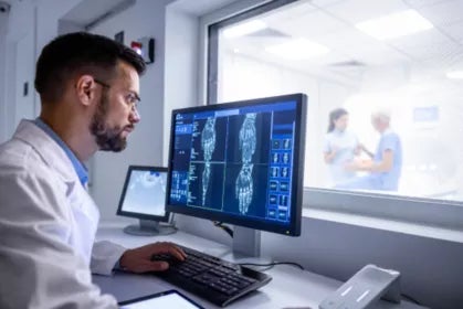
These technologies use X-rays, sound waves, magnetic fields or radioactive substances to create detailed images. Professionals specialising in this field, such as Senior Technicians in Diagnostic Imaging and Nuclear Medicine , are the ones who operate this equipment and obtain the images, which are then interpreted by radiologists.

Main types of imaging tests
X-rays (Radiography)
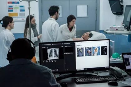
Radiography is perhaps the best-known of all radiological or diagnostic imaging tests. It uses a small dose of radiation to create a two-dimensional image of the inside of the body. These are incredibly useful for examining bones, detecting fractures or pneumonia, or identifying foreign objects in the body.
Ultrasound
Ultrasound is a safe, non-invasive technique that uses high-frequency sound waves to produce real-time images. Do you know how bats navigate in the dark? Ultrasound works in a similar way, sending out waves that bounce off organs and tissues to create an image on a screen. It is very popular during pregnancy for viewing the baby, and also for examining soft tissues such as the liver, kidneys and gallbladder.
Computed tomography (CT)
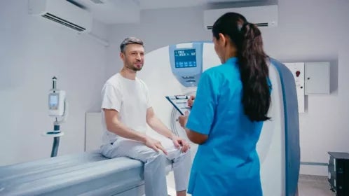
Also known as a CAT scan ( Computed Axial Tomography), this technique combines multiple X-rays taken from different angles to create a detailed three-dimensional image. It is as if the body were cut into thin virtual ‘slices’, allowing doctors to see the location and extent of tumours, internal bleeding or blood clots. A CT scan is much more detailed than a simple X-ray and is crucial in emergency situations.
Magnetic Resonance Imaging (MRI)
Magnetic resonance imaging does not use radiation. Instead, it uses a powerful magnetic field and radio waves to generate highly accurate images of organs and soft tissues, such as the brain, spinal cord, muscles and ligaments. Whilst it is not ideal for visualising bones, it is the test of choice for diagnosing sports injuries, neurological problems and joint disorders. Its high resolution allows it to detect abnormalities that other tests might overlook.
Nuclear medicine
In this type of test, a small amount of radioactive material (known as a radiopharmaceutical) is introduced into the body. Once inside, this material accumulates in a specific organ or type of tissue. A special camera, called a ‘ gamma camera ’, detects the radiation and creates an image showing how the organ is functioning. Unlike other techniques, which show the anatomy, nuclear medicine reveals the organ’s function, making it essential for the diagnosis and monitoring of diseases such as cancer and thyroid problems.
Radiological or diagnostic imaging tests: Clinical applications
Radiological or diagnostic imaging tests have virtually unlimited applications in medicine. They are used to:
- Detect fractures, dislocations and bone problems
- Diagnose heart and lung diseases
- Assess abdominal organs such as the liver, kidneys and pancreas
- Detect tumours and cancers at an early stage
- Monitor the progress of treatments
- Guide surgical procedures and biopsies
- To study foetal development during pregnancy
Examples across different medical specialities
Each medical speciality uses different types of imaging tests according to its specific needs:
· Orthopaedics: X-rays for fractures, MRI scans for ligament and cartilage injuries.
· Cardiology: Echocardiograms, angiograms and cardiac MRI scans.
· Neurology: CT scans and MRI scans of the brain and spinal cord.
· Oncology: PET-CT scans, MRI scans and CT scans to detect and monitor tumours.
· Gynaecology: Pelvic ultrasounds, mammograms and MRI scans.
Make a difference in people’s lives with UAX FP
Did you know that at UAX FP you can study for a degree in Diagnostic Imaging and Nuclear Medicine via their online programme? This course allows you to combine your studies with other commitments, learning from experts in the field through a practical and flexible approach.
If you see yourself in the future as a professional who makes a difference to people’s lives, helping doctors make accurate diagnoses and patients feel more confident, then this is your calling.
Do it at UAX!